
X-ray spectroscopy
In collaboration with Jacinto Sá (Uppsala University and Institute Physical Chemistry, Polish Academy of Sciences in Warsaw, PL) and Jakub Szlachetko (Institute of Nuclear Physics, Polish Academy of Sciences in Krakow, PL) ELI Beamlines RP4 is developing a setup for time-resolved X-ray spectroscopy at the laser-driven Plasma X-ray Source. The first generation setup will be a von-Hamos spectrometer for absorption spectroscopy. Future upgrades will allow simultaneous measurements using X-ray absorption and X-ray emission spectroscopy methods.
It is important to note, that X-ray absorption provides information about the unoccupied density of states of an scattering atom whereas X-ray emission spectroscopy reflects occupied density of states, and when applied together, providing the detailed picture of the molecular orbitals, giving insight into the oxidation state, spin state, and ligand environment around a specific absorbing atom. Furthermore, because of the hard X-ray light penetration, X-ray absorption and X-ray emission spectroscopies can be performed in ambient environments, thus the experimental conditions can be easily changed and controlled. The possibility of in situ experiments provides information about electronic structure under ambient/operational conditions, i.e., direct observation of species at the molecular level in low (biological) concentrations without the need for preconcentration, extraction or crystallization. In combination of X-ray absorption and X-ray emission methods with pulsed X-ray beam delivered at ELI will provide unique opportunity to study dynamics and mater transformations at femto-second time scales. For example, the setup will be used to determine charge dynamics around transition metals systems, which are of prime importance in catalysis, photosynthesis and light-activated cancer therapy.
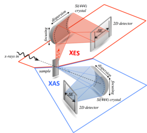
The conceptual design of a double von Hamos spectrometer set up for simultaneous emission and absorption spectroscopy.
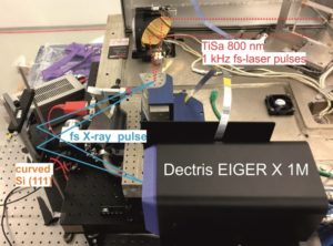
The first spectrometer prototype under evaluation at the ELI Beamlines facility in October 2017.
Two recent publications highlight the importance of complementary time-resolved X-ray and optical spectroscopy:
Transferring the entatic-state principle to copper photochemistry
Nature Chemistry volume 10, pages 355–362 (2018)
B Dicke, A Hoffmann, J Stanek, et al.
Structural dynamics upon photoexcitation-induced charge transfer in a dicopper(I)–disulfide complex
Phys. Chem. Chem. Phys., 20, 6274-6286 (2018)
Naumova, D. Khakhulin, M. Rebarz, et al.
Time-resolved X-ray diffraction (trXRD)
Since the discovery made by Max von Laue in 1912 of the diffraction of X-rays by crystals, the X-ray diffraction technique has played a strategic role in direct probing and in revealing the time-averaged static structure of matter. Because of these diffraction techniques, three-dimensional structures are now solvable with the atomic scale resolution in systems ranging from small molecules to crystals and from DNA and proteins to viruses and particles [1]. Diffraction provides a way of easily understanding why the properties of diamond are so different from those of graphite in spite of the fact that both diamond and graphite consist of the same chemical element of carbon. This is because diamond and graphite possess different special distributions of elementary carbon atoms as shown in the figure above.
However, a full description of phase transformation, chemical reactions, or biological functions requires a real-time visualization of the actual steps characterizing a given process. The time scales for our areas of interest can range from femtoseconds for molecular and phase transformations through picoseconds to the millisecond scale for visco-elastic flow and dissipation in liquids and gasses. One of the great dreams in science is to be able to create an “atomic/molecular movie” that permits the recording of motions as the causal events of interest. The aim is to observe the whole process of transition from an initial state/configuration of matter through intermediate states to a final state/configuration during a chemical reaction, a phase transformation, or any other type of dynamical process. Therefore, trXRD, which is often called ultrafast X-ray diffraction (UXRD) in the literature, has become more and more prevalent in various scientific disciplines that involve the direct observation of atomic motion in real time.
Typical trXRD experiments are carried out in a pump-probe mode. This method, suitable for studying quantitatively reproducible dynamics, relies on creating a time-delayed sequence of two short pulses: a “pump” pulse excites the matter from the balance and a “probe” pulse of X-rays is used to create or take ‘snapshots’ of the matter structure at a particular time during a dynamical response of the matter. It is then possible to trace out the structural response of a system as a function of time by repeatedly applying these two pulses to the matter with different relative delays.
The ELI Beamlines facility will develop different types of secondary X-ray sources. The X-ray diffraction beam line will rely on an X-ray plasma source driven by the laser system L1 with high average pulse power up to 100 mJ, a 1 kHz repetition pulse rate, and pulse duration of about 100 fs. The X-ray energy of spectra lines is expected to be from 5 to 10 keV. The part of the X-rays that was originally illuminated into a whole 4π space will be collected using suitable optics. The X-rays that are collected will be focused on the sample situated in the middle of a goniometer. The expected intensity on the sample should be about 109 photons/s with a spot size of a few tens to a few hundreds µm2. Diffracted intensity will be detected by a two-dimensional detector working in a single photon counting mode. One great advantage of the fact that trXRD uses a laser-driven X-ray source will be near-perfect synchronization of the temporal overlap of the pump laser pulses and X-ray probe pulses (low-jitter).
Time-resolved X-ray spectroscopy
The ultrafast pump-probe X-ray Absorption and Fluorescence Spectroscopy will be used to capture the photo-initiated chemical dynamics. Using either the Plasma X-ray Source (PXS) or Betatron source, K- and L-edge spectroscopy will be realized by imaging into the sample X-rays with photon energies in the range of ten keV and hundreds of eV, respectively. The X-ray sources will emit, depending on the type and driving laser conditions, suitable broad bandwidth X-radiation from the soft to hard X-ray regime. The polychromatic X-ray emission will be captured and focused into the optically pumped interaction region of the sample using a fiber-optic X-ray lens. After transmission through the sample solution, the X-ray radiation will be spectrally dispersed and detected by an X-ray CCD camera or Dectris Eiger X pixel array detector. Owing to the ultrashort X-ray pulse length, measurements of structural dynamics will be possible with a temporal resolution of 300 fs. The liquid samples will be continuously pumped through a fused silica capillary with about 500 μm inner diameter, or through a microfluidic nozzle creating a flat sheet jet.
REFERENCES:
[1] A.H. Zewail. “The new age of structural dynamics,” Acta Crystallographica A 66, 135-136 (2010).
Petr Bruza
Štefan Michalik (now at Diamond Light Source)